Testing Plant Substances as Potential Medicines
Purpose: We will find what plant materials, that are native, contains elements that will help bacteria grow.
Materials:
Materials:
- Balance, weigh boat, lab scoops
- LB broth base
- Media bottles, 250 mL
- Sterilizer/autoclave
- Water bath, 37*C, shaking
- Sterile LB agar
- Laminar flow hood and disinfectant
- Plastic safety glasses
- Bunsen burner and gas lighter
- Inoculating loop, Ni/Cr wire
- Petri dishes, 60x15mm, sterile
- E. coli JM109 (stock plate)
- Plant specimen
- Mortar and pestle
- Pipet, 10 mL and pump
- Plastic funnels, short-stemmed
- Filter paper disks, 5mm diameter
- 100 mL beakers
- Syringe, 10 mL and filter, 0.2 micrometers
- Reaction tubes and rack, 1.7 mL
- Methanol, absolute
- Pipet, 1 mL and pump
- Dry block heater/heat block
- Forceps, fine-tipped
- Ampicillin
- Glass spreader
- Incubator oven, 37*c
Procedure:
Part 2
4. Grind 2 g of plant tissue (leaves) with 10 mL of deionized water in mortar and pestle, and let sit for 3 minutes. Channel in 11 cm pipe, sanitize extricate with syringe channel, and gather 1 mL of concentrate in marked 1.7 mL microtube.
5. Rehash Step 4, with the exception of supplant deionized water with methanol. Place 1.7 mL tube with 1 mL of methanol concentrate in 65*C warmth piece (tops open) for 24 hours to vanish methanol. Reconstitute dry matter in microtube with 1 mL of deionized water.
6. Rehash Step 4 and 5 for six examples and mark them.
7. Drop channel paper circles in every sifted concentrate tube utilizing sterile forceps (disinfected by being flared in liquor).
8. Set up three negative control circles of just methanol and sterile and refined water.
9. Set up six positive control plates of ampicillin arrangement.
10. Permit plates to be soaked with the concentrate (maybe overnight).
11. Close tubes. Store all specimens at 4*C until prepared to utilize.
Part 3
12. Use a clean pipet to move 1 ml of E.coli broth to the middle of a petri dish. sterilize a spreading loop with fire and alcohol, to spread the bacteria evenly. Cover, and leave for at least 15 minutes.
13. Using forceps, place 1 dish in each quadrant, 2 cm from the edge of the petri dish. Place the methanol samples in one dish and the water samples on another.
14. Repeat step 13, in order to end up with 3 methanol and 3 water replicates.
15. Place a negative control disk in the center of the appropriate plate. Then a positive control with amplicillin in a quadrant on each plate.
16. In the end you should have 6 petri dishes with a negative control in the middle and a a positve control and three sample disks.
17. Guarantee that the disk hold fast to surface of agar. Alter the plates and brood at 37*C for 24 to 48 hours.
18. After incubation, search for at the plates with plant concentrate circles for zones of inhibition, clear rasnge shaped by inhibitory (abatement in real life) activity of a substance in the plant material around the circle. Photo the plates, marking any restraint of bacterial development.
19. Make an information table for the repeats and midpoints. Incorporate depictions of the bacterial garden around every circle. Record the breadth and clarity of any cleared zones around the circles in quantitative estimations.
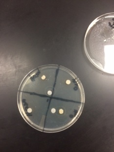
Results
The plant samples I used had a negative result. The methanol disc had larger rings around it (1.1 cm), which seemed to have killed off some bacteria, but had a negative result due to the disk had some bacteria up against it.There are still signs of bacterial activity around the inner rims, menaing that the disc did not kill off any bacteria but instead pushed the bacteria farther out. The plant sample that had been doused in water clearly provided negative results. These disks were only 0.1-0.2 in width, and were most likely due to water.
Data Analysis
In the end none of the extracts gave me a positive outcome. My plant doesn't affect the E.coli bacteria. The controls turned out how we expected them. There is a possibility that error occurred due to being not accurate in placing them, and misplacing disks in wrong areas. I also had the negative and positive control slide into different areas over night. This can cause area in my results. Due to negative results I would use different solvents in the futrue. Also, due to the movement of my disk over night, I the future I would let the bacteria soak in more, to insure my data isn't read wrong. For the next steps i would test a new plant since this plant did not have any positive outcomes.
Thinking Like a Bio Technician Questions
- We only tested our extract for E.coli, this means it could be antimicrobial for a different bacteria. So it is possible that the extract is an antimicrobial agent.
- It is a problem because you do not want the alcohol to tamper with the extract and have us test the alcohol instead. The alcohol could kill the bacteria as well.
- Through chromatography we can figure out what compound is causing microbial action. Each compound can have more than one compound in it so through this use we can identify it.